Multimodal Coherent Imaging of Retinal Biomarkers of Alzheimer's Disease

Alzheimer’s disease (AD) is a neurodegenerative disease that currently affects 5.8 million Americans. Important efforts have been made to clinically diagnose AD early, which involves neuroimaging techniques to extract correlated pathological changes in the brain. However, the invasiveness and high-cost nature of the neuroimaging techniques have limited their applicability on a population scale. Many researchers have therefore shifted to study AD noninvasively through the retina, which is an extension of the central nervous system that also manifests pathological changes from AD. The use of optical retinal imaging tools, therefore, has become a promising approach that is more accessible and increasingly low cost.
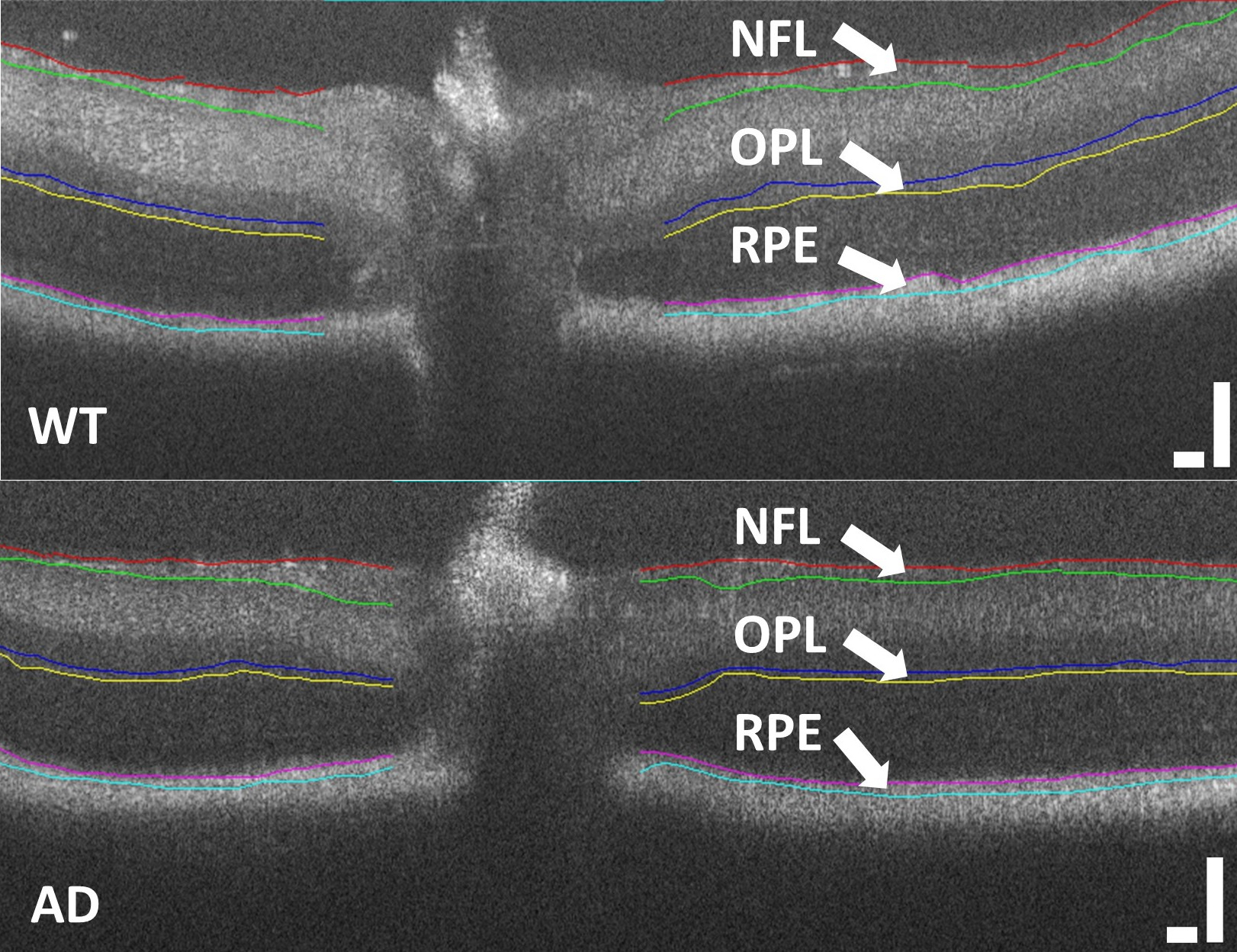
Our lab aims to develop low-cost retinal imaging tools to extract quantitative changes in the retina for AD. Specifically, optical coherence tomography (OCT) is used as image guidance for angle-resolved low-coherence interferometry (a/LCI) to make light scattering measurements on the retina. For OCT, we use low-cost OCT to overcome the costly and bulky nature of current commercial systems, making the technology more accessible as a screening tool for AD. In addition to morphological information from OCT, we also improve the diagnostic power of AD correlated retinal parameters with additional light scattering measurements from a/LCI. Our previous studies in the transgenic AD mouse models have demonstrated the potential of a combined OCT and a/LCI retinal imaging system to extract AD biomarkers in future clinical studies. Figure from Song et. al. 2020.