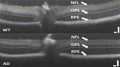
Our lab aims to develop robust clinical imaging tools to extract quantitative changes in relevant biomarkers of disease, such as in Alzheimer’s disease.
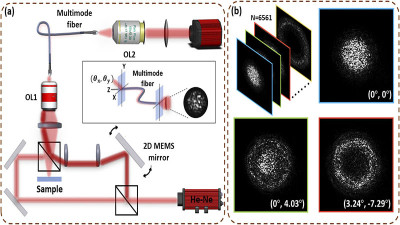
We are currently developing an optomechanical system to achieve fast collection and reconstruction of angular scattering distributions using a single multimode fiber.
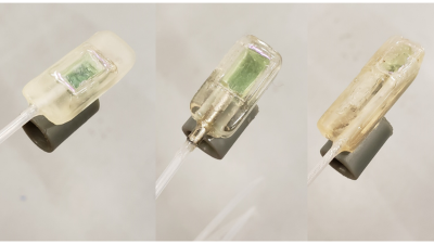
We are now integrating a/LCI technology with our recently developed 3D printed paddle probes to provide a reading on dysplasia, as well as OCT images of Barrett’s esophagus, during a single endoscopic procedure.
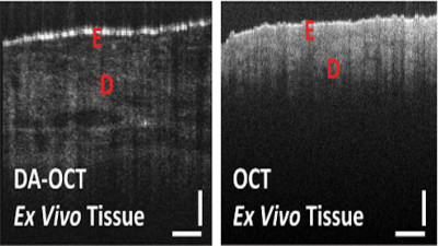
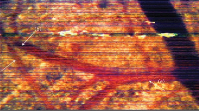
DA-OCT exposes morphology otherwise undetected using conventional OCT using a distinct illumination and collection scheme to triangulate the signal to layers deep beneath the tissue surface.
By isolating narrowband regions of an OCT interferogram, spectroscopic optical coherence tomography (SOCT) allows for a more comprehensive assessment of tissues without additional necessary hardware.
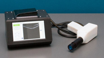
We seek to minimize the cost of optical coherence tomography (OCT) and provide comparable performance with commercial clinical systems by using inexpensive components such as uncooled SLD for the light source and liquid lens for the scanner.
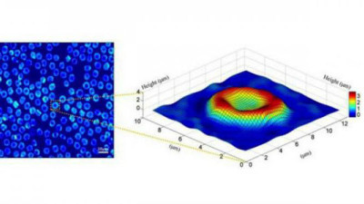
Quantitative phase cytometry is an effective label-free research tool used for high throughput examination of spatial and temporal properties of biological samples, by measuring the optical path delays of light.
We are developing several methods to utilize quantitative phase imaging (QPI) for molecular, structural, and mechanical characterization of cells, including cancer cells and red blood cells (RBCs).